Skin Type: 2

  • Alopecia Areata

    Alopecia Areata

    Alopecia Areata

    Definition: A common autoimmune condition that is marked by abrupt, non-scarring hair loss on the scalp and other parts of the body go (1)

    Etiology: Many etiologies have been proposed but it is believed that the condition is caused by an autoimmune attack on the hair follicles, leading to hair loss. In addition, environmental factors, genetic predisposition and stress may also play a role (1,2).

    Epidemiology: Alopecia Areat (AA) affects 0.1-0.2% of the overall population with no gender predilection. (1,2).

    Signs: AA is characterized by well-demarcated oval or round patches of hair loss, the lesions may be solitary or multiple with 90% of cases affecting the scalp (2).

    Symptoms: Typically asymptomatic however some patients may experience nail changes, tingling or burning sensation, irritated eyes if loss of hair in eyelashes or eyebrows (3).

    Differentials: Telogen effluvium, Trichotillomania,Tinea capitis, Early scarring Alopecia, Anagen effluvium, Systemic lupus erythematosus, Secondary syphilis and androgenic Alopecia (1,2). 

    Diagnosis: The diagnosis is clinical with a thorough history and routine investigations such as complete hemogram, anemia panel, erythrocyte sedimentation rate, etc (2). Dermoscopic histological examination is a great tool in diagnosing AA and in more uncertain cases a biopsy may also be performed (1).

    Treatment: In a majority of cases the condition is self-limited and regrowth typically will happen within a year (1). In patients where AA is persistent corticosteroids can be prescribed to stimulate hair growth follow by minoxidil to maintain the growth (3).

    References: (AMA)

    1.     Khan Mohammad Beigi P. Alopecia areata. Published online 2018. doi:10.1007/978-3-319-72134-7 

    1. Amin SS, Sachdeva S. Alopecia areata: A Review. Journal of the Saudi Society of Dermatology & Dermatologic Surgery. 2013;17(2):37-45. doi:10.1016/j.jssdds.2013.05.004 
    2. Ludmann P. Hair loss types: Alopecia areata diagnosis and treatment. American Academy of Dermatology. 2023. Accessed August 18, 2024. https://www.aad.org/public/diseases/hair-loss/types/alopecia/treatment. 
  • Allergic Contact Dermatitis

    Allergic Contact Dermatitis

    Allergic Contact Dermatitis 

    Definition: Allergic contact dermatitis (ACD) is a skin condition that occurs when the skin comes into contact with an allergen, leading to an immune response and subsequent inflammation(1).

    Etiology: ACD is caused by an allergic reaction to substances that come into direct contact with the skin. Common allergens include nickel, fragrances, preservatives, latex, and certain plants like poison ivy (1,3). The immune system recognizes these substances as harmful, triggering an inflammatory response(1).

    Epidemiology: ACD affects individuals of all ages, but it is more prevalent in adults due to cumulative exposure to allergens over time. It is common with some studies demonstrating a prevalence rate of 20% in the general population (2). It is particularly common in certain occupations that involve frequent contact with irritants and allergens, such as healthcare workers, hairdressers, and construction workers (2). 

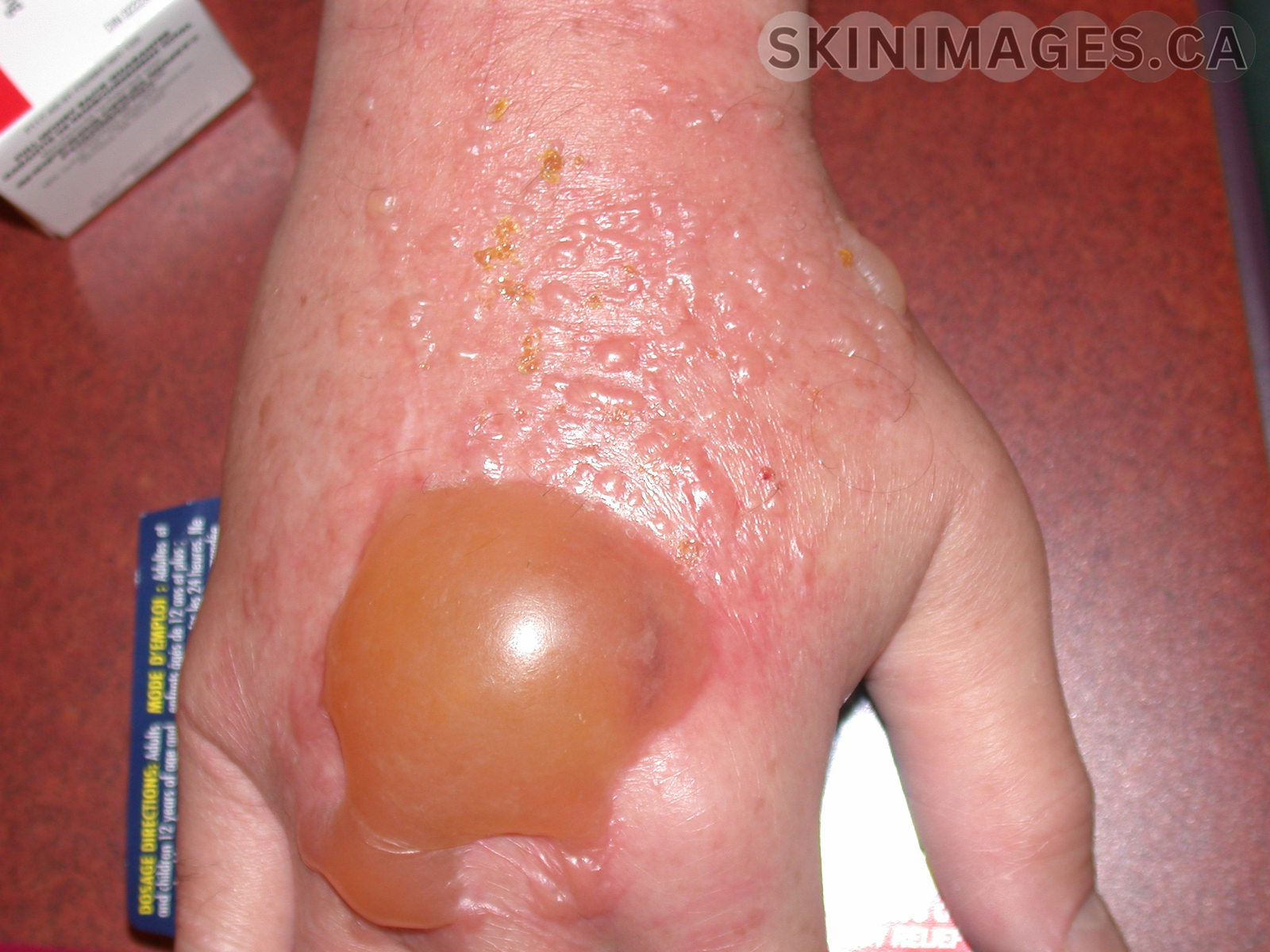
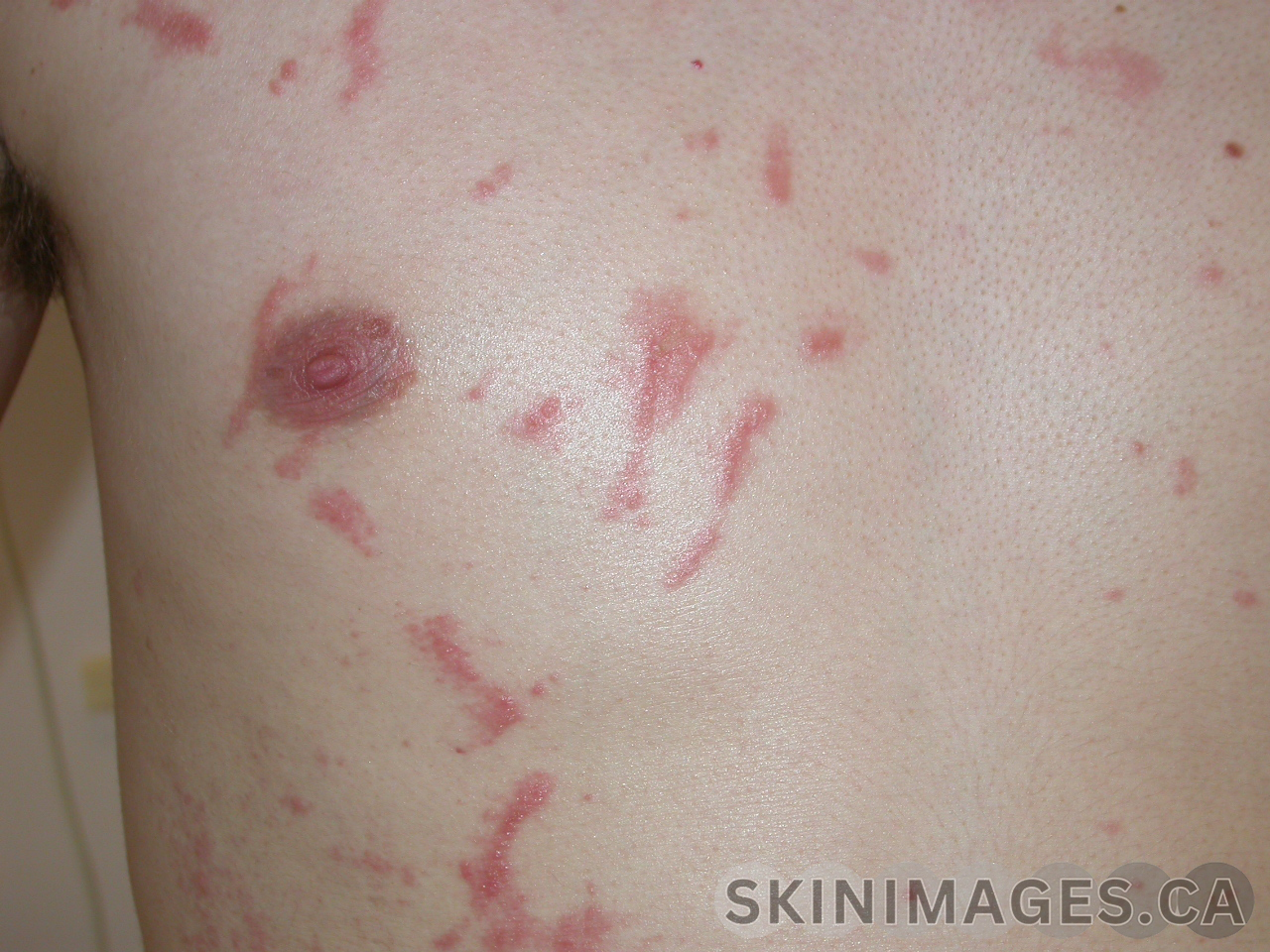
    Signs: Key signs of ACD include erythema, edema, vesicles, and lichenification in chronic cases (1). These signs typically appear in areas directly exposed to the allergen(1,2).

    Symptoms: Symptoms of ACD include intense itching, burning, and discomfort at the site of contact(3). The skin may also become dry, cracked, and scaly if exposure continues or if the dermatitis becomes chronic(3)

    Differentials: Differential diagnoses for ACD include irritant contact dermatitis, atopic dermatitis, psoriasis, fungal infections, and seborrheic dermatitis (1).

    Diagnosis: Diagnosis of ACD is primarily clinical, based on the history of exposure and characteristic skin findings(1). Patch testing is the gold standard and is used to identify the allergens that cause allergic contact dermatitis (1).

    Treatment: The cornerstone of ACD treatment is identifying and avoiding the offending allergen(1). Topical corticosteroids are commonly prescribed to reduce inflammation and alleviate symptoms(1). In severe cases, systemic corticosteroids may be required. Avoidance necessitates thorough label-checking for the allergen and related cross-reactive substances. Understanding these cross-reactive ingredients and knowing where a particular allergen is commonly found can aid in effectively advising the patient (1).

    References: (AMA)

    1. Kanwaljit K. Brar MD, AbstractObjectiveTo familiarize the reader with the mechanisms and causes of contact dermatitis.Data SourcesRecent research articles, Dhingra N, et al. A review of contact dermatitis. Annals of Allergy, Asthma & Immunology. October 20, 2020. Accessed August 10, 2024. https://www.sciencedirect.com/science/article/pii/S1081120620310802.
    2. Stacy Nassau MD, Jacob SE, Warshaw EM, et al. Allergic contact dermatitis. Medical Clinics of North America. October 28, 2019. Accessed August 10, 2024. https://www.sciencedirect.com/science/article/pii/S0025712519300884. 
    3. Brandon L. Adler M. Allergic contact dermatitis. JAMA Dermatology. March 1, 2021. Accessed August 10, 2024. https://jamanetwork.com/journals/jamadermatology/article-abstract/2775575. 
  • Acute Generalized Exanthematous Pustulosis (AGEP)

    Acute Generalized Exanthematous Pustulosis (AGEP)

    Acute Generalized Exanthematous Pustulosis (AGEP)

    Definition:  Acute Generalized Exanthematous Pustulosis (AGEP) is a severe cutaneous adverse reaction characterized by the abrupt appearance of numerous nonfollicular, sterile pustules on an erythematous base (1).

    Etiology: AGEP is commonly triggered by medications, particularly antibiotics and antifungals, but can also be associated with infections and other factors(1). It is most commonly triggered by the following drugs: pristinamycin,aminopenicillins, quinolones, hydroxychloroquine, sulfonamides, terbinafine, diltiazem, ketoconazole, and fluconazole(1,2)

    Epidemiology: AGEP is a rare condition, with an estimated incidence ranging from 1 to 5 cases per million people (2).

    Signs: Key signs include widespread pustular rash and fever, with the pustules favoring the trunk and intertriginous regions. Leukocytosis with an elevated neutrophil count is also a feature (1). The most frequent features in patients with systemic involvement are liver, kidney, and lung dysfunction (1,3).

    Symptoms: Patients may experience fever, chills, and systemic malaise alongside the skin manifestations(3).

    Differentials: Differential diagnoses include pustular psoriasis, generalized pustular exanthema, and drug-induced eruptions (1,3).

    Diagnosis: Diagnosis is based on both clinical presentation and histological criteria. The EuroSCAR group created a standardized AGEP validation score that classifies patients into categories of definite, probable, possible, or no AGEP, based on morphology, clinical progression, and histology.

    Treatment: Management involves discontinuation of the offending drug, supportive care, and, in severe cases, systemic corticosteroids (1,2). During the pustular phase, moist dressings and antiseptic solutions can be used to prevent infection (1).

    References: (AMA)

    1. Szatkowski J, Schwartz RA. Acute generalized exanthematous pustulosis (AGEP): A Review and update. Journal of the American Academy of Dermatology. 2015;73(5):843-848. doi:10.1016/j.jaad.2015.07.017
    2. Oh DA, Yeo YW, Choo KJ, Pang SM, Oh CC, Lee HY. Acute generalized exanthematous pustulosis: Epidemiology, clinical course, and treatment outcomes of patients treated in an asian academic medical center. JAAD International. 2021;3:1-6. doi:10.1016/j.jdin.2020.12.004 
    3. Parisi R, Shah H, Navarini AA, et al. Acute generalized exanthematous pustulosis: Clinical features, differential diagnosis, and management. American Journal of Clinical Dermatology. 2023;24(4):557-575. doi:10.1007/s40257-023-00779-3 
  • Acute Generalized Exanthematous Pustolosis (AGEP)

    Acute Generalized Exanthematous Pustolosis (AGEP)

    Acute Generalized Exanthematous Pustulosis (AGEP)

    Definition:  Acute Generalized Exanthematous Pustulosis (AGEP) is a severe cutaneous adverse reaction characterized by the abrupt appearance of numerous nonfollicular, sterile pustules on an erythematous base (1).

    Etiology: AGEP is commonly triggered by medications, particularly antibiotics and antifungals, but can also be associated with infections and other factors(1). It is most commonly triggered by the following drugs: pristinamycin,aminopenicillins, quinolones, hydroxychloroquine, sulfonamides, terbinafine, diltiazem, ketoconazole, and fluconazole(1,2)

    Epidemiology: AGEP is a rare condition, with an estimated incidence ranging from 1 to 5 cases per million people (2).

    Signs: Key signs include widespread pustular rash and fever, with the pustules favoring the trunk and intertriginous regions. Leukocytosis with an elevated neutrophil count is also a feature (1). The most frequent features in patients with systemic involvement are liver, kidney, and lung dysfunction (1,3).

    Symptoms: Patients may experience fever, chills, and systemic malaise alongside the skin manifestations(3).

    Differentials: Differential diagnoses include pustular psoriasis, generalized pustular exanthema, and drug-induced eruptions (1,3).

    Diagnosis: Diagnosis is based on both clinical presentation and histological criteria. The EuroSCAR group created a standardized AGEP validation score that classifies patients into categories of definite, probable, possible, or no AGEP, based on morphology, clinical progression, and histology.

    Treatment: Management involves discontinuation of the offending drug, supportive care, and, in severe cases, systemic corticosteroids (1,2). During the pustular phase, moist dressings and antiseptic solutions can be used to prevent infection (1).

    References: (AMA)

    1. Szatkowski J, Schwartz RA. Acute generalized exanthematous pustulosis (AGEP): A Review and update. Journal of the American Academy of Dermatology. 2015;73(5):843-848. doi:10.1016/j.jaad.2015.07.017
    2. Oh DA, Yeo YW, Choo KJ, Pang SM, Oh CC, Lee HY. Acute generalized exanthematous pustulosis: Epidemiology, clinical course, and treatment outcomes of patients treated in an asian academic medical center. JAAD International. 2021;3:1-6. doi:10.1016/j.jdin.2020.12.004 
    3. Parisi R, Shah H, Navarini AA, et al. Acute generalized exanthematous pustulosis: Clinical features, differential diagnosis, and management. American Journal of Clinical Dermatology. 2023;24(4):557-575. doi:10.1007/s40257-023-00779-3