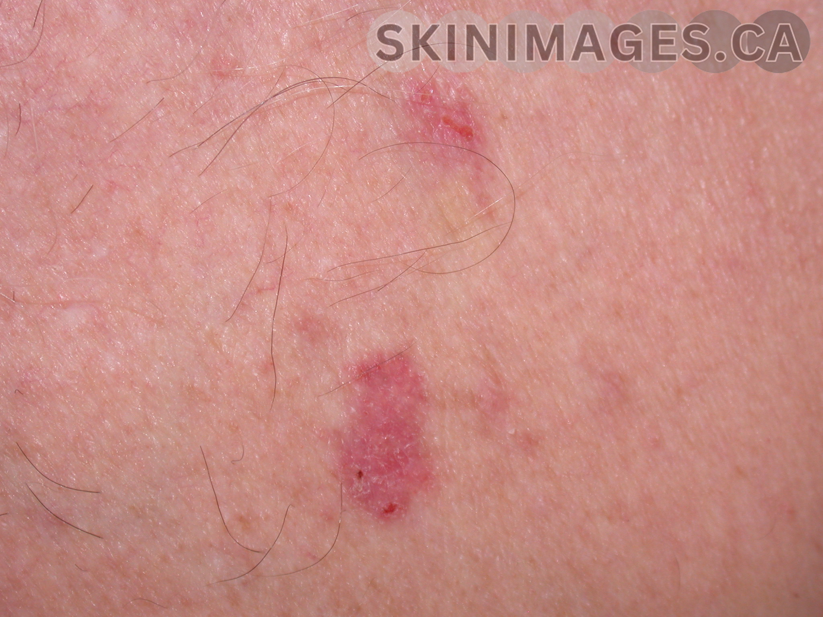
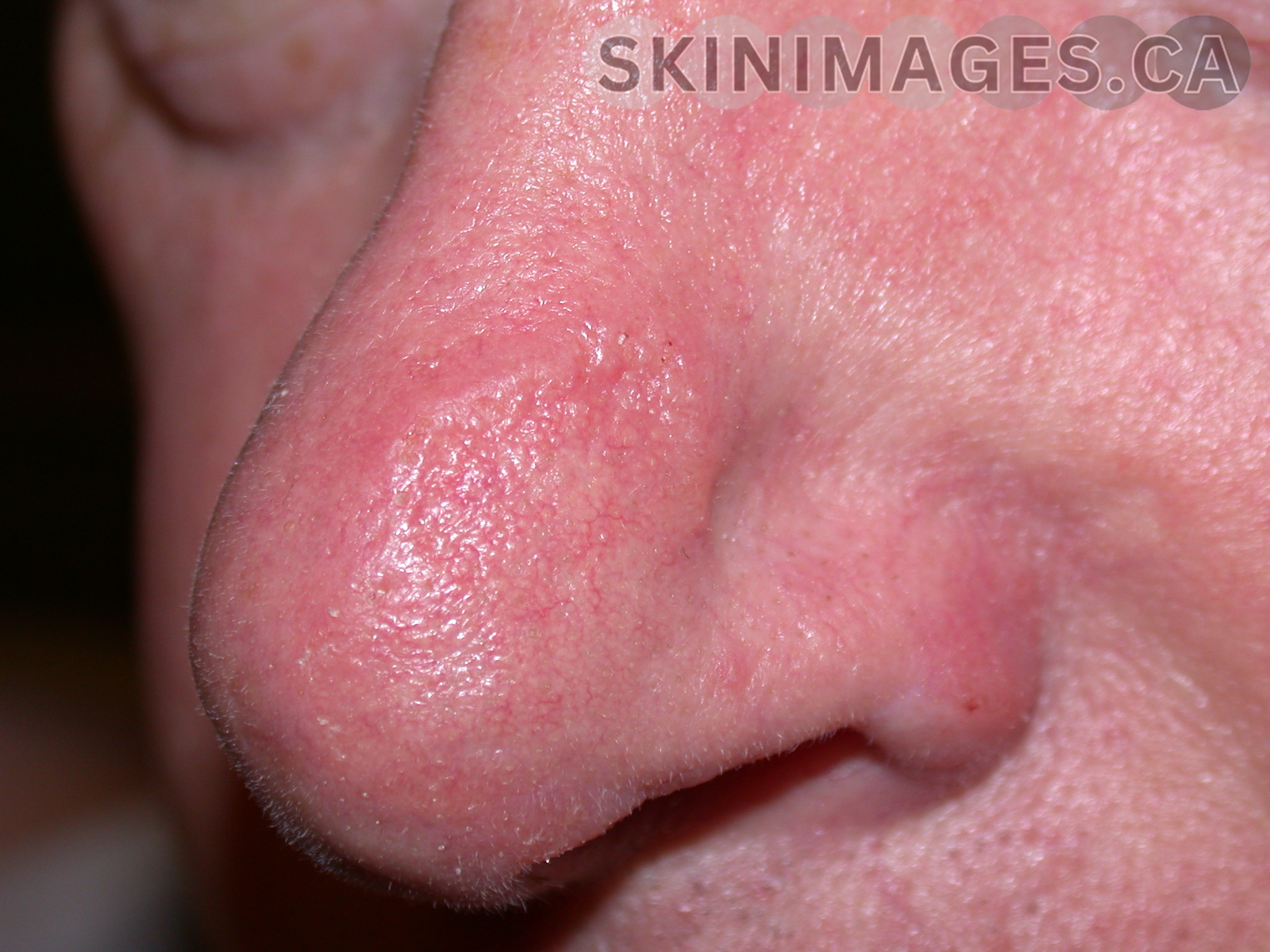
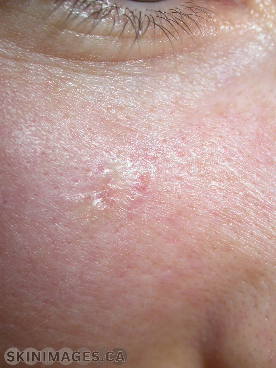
Skin Type: 2

  • Folliculitis Decalvans

    Folliculitis Decalvans

    Folliculitis Decalvans

    Condition Name: Folliculitis decalvans

    Definition: Folliculitis decalvans is a chronic inflammatory disorder that primarily affects hair follicles, leading to scarring alopecia. It is characterized by recurrent pustules and crusts around hair follicles, eventually causing permanent hair loss in the affected areas.

    Etiology: The etiology of folliculitis decalvans is not fully understood, but it is believed to involve bacterial infection, particularly with Staphylococcus aureus, combined with an abnormal immune response.

    Epidemiology: Folliculitis decalvans is relatively rare and typically affects adults, with a slight male predominance. The exact prevalence is not well documented.

    Signs: Clinically, it presents with tufted folliculitis, where multiple hairs emerge from a single follicular opening surrounded by inflammation, along with recurrent pustules and crusts on the scalp.

    Symptoms: Symptoms include itching, pain, and the presence of pustules and crusts on the scalp.

    Differentials: Conditions such as dissecting cellulitis, lichen planopilaris, and acne keloidalis nuchae should be considered.

    Diagnosis: Diagnosis is based on clinical examination, patient history, and, if needed, biopsy to confirm the presence of inflammation and scarring around hair follicles.

    Treatment: Treatment focuses on controlling infection and inflammation to prevent further hair loss. Topical and systemic antibiotics, such as clindamycin and rifampicin, are commonly used. Additionally, anti-inflammatory medications like corticosteroids and immunosuppressive agents may be prescribed. Early intervention is crucial to manage symptoms and slow the progression of scarring.

    References:

    ●       Scheinfeld, N. (2003). “Folliculitis decalvans: A review of the literature.” American Journal of Clinical Dermatology, 4(6), 369-374. doi:10.2165/00128071-200304060-00003

    ●       Otberg, N., & Shapiro, J. (2008). “Diagnosis and management of primary cicatricial alopecia: Part II. Treatment.” Journal of the American Academy of Dermatology, 59(1), 1-22. doi:10.1016/j.jaad.2008.01.072

  • Basal Cell Carcinoma – Nodular

    Basal Cell Carcinoma – Nodular

    Nodular Basal Cell Carcinoma 

    Definition: Nodular Basal Cell carcinoma is a subtype of BCC that is marked by raised, pearly flesh coloured nodules with melanogenesis typically localized to the face (2,3).

    Etiology: Similar to other BCCs, Nodular BCC is caused primarily by exposure to ultraviolet light as well as genetic factors (3). In the skin of color, pigmented Nodular BCCs are more common (1).

    Epidemiology: Nodular Basal Cell carcinoma is the most common subtype of BCC and has a high incidence in individuals with fair skin with a significant history of sun exposure (1). Approximately 60% to 80% of BCC are of the Nodular subtype (4).

    Signs: Nodular BCC manifests as skin-coloured papules or nodules with pearly or translucent telangiectatic border (3,4). It may be described as having a rolled border in which the edges of the lesion are higher than the center (4). Large lesions may include ulceration (4).

    Symptoms: Typically asymptomatic but with time the lesions may crust or bleed (2,3).

    Differentials: Trichoblastoma and trichoepithelioma (1).

    Diagnosis: Diagnosis is confirmed through a shave or punch biopsy but clinical exam and dermoscopy can supplement the assessment (2).

    Treatment: Treatment is usually surgical involving either surgical excision or Mohs micrographic surgery (1,3). Non-surgical options include cryotherapy, topical therapies or radiation in some cases (1,3).

    References: (AMA)

    1.      1. McDaniel B. Basal cell carcinoma. StatPearls [Internet]. March 13, 2024. Accessed August 18, 2024. https://www.ncbi.nlm.nih.gov/books/NBK482439/#:~:text=BCC%20typically%20presents%20as%20a,rolled%20or%20rodent%20ulcer%20appearance.

    2.     Oakley A. Basal cell carcinoma: Symptoms, causes, and treatment – dermnet. DermNet®. July 3, 2024. Accessed August 18, 2024. https://dermnetnz.org/topics/basal-cell-carcinoma. 

    3.     1. Robert S Bader M. Basal cell carcinoma. Practice Essentials, Background, Pathophysiology. April 3, 2024. Accessed August 18, 2024. https://emedicine.medscape.com/article/276624-overview?gad_source=1&gbraid=0AAAAADoSQiUCwFng3uMBdxWlXHVuNMVrw&gclid=Cj0KCQjwt4a2BhD6ARIsALgH7Dqhf-P79uzm848PdW5k8auVX_QjoRGo38V0m2dFrNN42l3MpFCbyS0aAm4_EALw_wcB&form=fpf#a2.

    4.     Tanese K. Diagnosis and management of basal cell carcinoma. Current Treatment Options in Oncology. 2019;20(2). doi:10.1007/s11864-019-0610-0 

  • Basa Cell Carcinoma – Nodular

    Basa Cell Carcinoma – Nodular

    Nodular Basal Cell Carcinoma 

    Definition: Nodular Basal Cell carcinoma is a subtype of BCC that is marked by raised, pearly flesh coloured nodules with melanogenesis typically localized to the face (2,3).

    Etiology: Similar to other BCCs, Nodular BCC is caused primarily by exposure to ultraviolet light as well as genetic factors (3). In the skin of color, pigmented Nodular BCCs are more common (1).

    Epidemiology: Nodular Basal Cell carcinoma is the most common subtype of BCC and has a high incidence in individuals with fair skin with a significant history of sun exposure (1). Approximately 60% to 80% of BCC are of the Nodular subtype (4).

    Signs: Nodular BCC manifests as skin-coloured papules or nodules with pearly or translucent telangiectatic border (3,4). It may be described as having a rolled border in which the edges of the lesion are higher than the center (4). Large lesions may include ulceration (4).

    Symptoms: Typically asymptomatic but with time the lesions may crust or bleed (2,3).

    Differentials: Trichoblastoma and trichoepithelioma (1).

    Diagnosis: Diagnosis is confirmed through a shave or punch biopsy but clinical exam and dermoscopy can supplement the assessment (2).

    Treatment: Treatment is usually surgical involving either surgical excision or Mohs micrographic surgery (1,3). Non-surgical options include cryotherapy, topical therapies or radiation in some cases (1,3).

    References: (AMA)

    1.      1. McDaniel B. Basal cell carcinoma. StatPearls [Internet]. March 13, 2024. Accessed August 18, 2024. https://www.ncbi.nlm.nih.gov/books/NBK482439/#:~:text=BCC%20typically%20presents%20as%20a,rolled%20or%20rodent%20ulcer%20appearance.

    2.     Oakley A. Basal cell carcinoma: Symptoms, causes, and treatment – dermnet. DermNet®. July 3, 2024. Accessed August 18, 2024. https://dermnetnz.org/topics/basal-cell-carcinoma. 

    3.     1. Robert S Bader M. Basal cell carcinoma. Practice Essentials, Background, Pathophysiology. April 3, 2024. Accessed August 18, 2024. https://emedicine.medscape.com/article/276624-overview?gad_source=1&gbraid=0AAAAADoSQiUCwFng3uMBdxWlXHVuNMVrw&gclid=Cj0KCQjwt4a2BhD6ARIsALgH7Dqhf-P79uzm848PdW5k8auVX_QjoRGo38V0m2dFrNN42l3MpFCbyS0aAm4_EALw_wcB&form=fpf#a2.

    4.     Tanese K. Diagnosis and management of basal cell carcinoma. Current Treatment Options in Oncology. 2019;20(2). doi:10.1007/s11864-019-0610-0 

  • Basal Cell Carcinoma – Nodular

    Basal Cell Carcinoma – Nodular

    Nodular Basal Cell Carcinoma 

    Definition: Nodular Basal Cell carcinoma is a subtype of BCC that is marked by raised, pearly flesh coloured nodules with melanogenesis typically localized to the face (2,3).

    Etiology: Similar to other BCCs, Nodular BCC is caused primarily by exposure to ultraviolet light as well as genetic factors (3). In the skin of color, pigmented Nodular BCCs are more common (1).

    Epidemiology: Nodular Basal Cell carcinoma is the most common subtype of BCC and has a high incidence in individuals with fair skin with a significant history of sun exposure (1). Approximately 60% to 80% of BCC are of the Nodular subtype (4).

    Signs: Nodular BCC manifests as skin-coloured papules or nodules with pearly or translucent telangiectatic border (3,4). It may be described as having a rolled border in which the edges of the lesion are higher than the center (4). Large lesions may include ulceration (4).

    Symptoms: Typically asymptomatic but with time the lesions may crust or bleed (2,3).

    Differentials: Trichoblastoma and trichoepithelioma (1).

    Diagnosis: Diagnosis is confirmed through a shave or punch biopsy but clinical exam and dermoscopy can supplement the assessment (2).

    Treatment: Treatment is usually surgical involving either surgical excision or Mohs micrographic surgery (1,3). Non-surgical options include cryotherapy, topical therapies or radiation in some cases (1,3).

    References: (AMA)

    1.      1. McDaniel B. Basal cell carcinoma. StatPearls [Internet]. March 13, 2024. Accessed August 18, 2024. https://www.ncbi.nlm.nih.gov/books/NBK482439/#:~:text=BCC%20typically%20presents%20as%20a,rolled%20or%20rodent%20ulcer%20appearance.

    2.     Oakley A. Basal cell carcinoma: Symptoms, causes, and treatment – dermnet. DermNet®. July 3, 2024. Accessed August 18, 2024. https://dermnetnz.org/topics/basal-cell-carcinoma. 

    3.     1. Robert S Bader M. Basal cell carcinoma. Practice Essentials, Background, Pathophysiology. April 3, 2024. Accessed August 18, 2024. https://emedicine.medscape.com/article/276624-overview?gad_source=1&gbraid=0AAAAADoSQiUCwFng3uMBdxWlXHVuNMVrw&gclid=Cj0KCQjwt4a2BhD6ARIsALgH7Dqhf-P79uzm848PdW5k8auVX_QjoRGo38V0m2dFrNN42l3MpFCbyS0aAm4_EALw_wcB&form=fpf#a2.

    4.     Tanese K. Diagnosis and management of basal cell carcinoma. Current Treatment Options in Oncology. 2019;20(2). doi:10.1007/s11864-019-0610-0 

  • Atopic Dermatitis

    Atopic Dermatitis

    Atopic Dermatitis 

    Definition: Atopic Dermatitis (AD) is a chronic inflammatory skin disorder marked by scaly, erythematous, and itchy skin lesions that are frequently seen in the body’s flexural surfaces (1).

    Etiology: Atopic dermatitis can often present as part of an atopic/allergic triad presenting with asthma and allergic rhinitis (1). Most individuals with AD will have a personal or family history and/or an increased production of immunoglobulin (Ig) E antibodies (4).

    Epidemiology: The onset is typically before the age of two with a reported 10% of cases presenting after the age of five (1). The prevalence in children is approximately 20% and in adults it ranges from 7% to 14% (2).

    Signs: Scaly, erythematous plaques usually accompanied by lichenification (4). 

    Symptoms: Patients present with dry skin and an itchy rash which can lead to infection and substantial reduction of quality of life (2).

    Differentials: Candidiasis, conditions that cause generalized pruritus, contact dermatitis, dermatitis herpetiformis, impetigo, lichen simplex chronicus, nummular eczema, psoriasis, scabies, seborrheic dermatitis, urticaria and xerosis (1).

    Diagnosis: Diagnosis is based on a comprehensive history and physical exam (1). If needed a biopsy or patch can be performed (3).

    Treatment: The goal of treatment is to relieve symptoms and increase the period in between flare-ups (1). Emollients are recommended as treatment with topical corticosteroids being first line treatment for flare-ups of atopic dermatitis (1).

    References: (AMA)

    1.      1. Berke R, Singh A, Guralnick M. Atopic dermatitis: An overview. American Family Physician. July 1, 2012. Accessed August 18, 2024. https://www.aafp.org/pubs/afp/issues/2012/0701/p35.html#clinical-presentation-and-differential-diagnosis.

    2.     Bylund S, Kobyletzki L, Svalstedt M, Svensson. Prevalence and incidence of atopic dermatitis: A systematic review. Acta Dermato Venereologica. 2020;100(12). doi:10.2340/00015555-3510 

    3.     Sidbury R, Alikhan A, Bercovitch L, et al. Guidelines of care for the management of atopic dermatitis in adults with topical therapies. Journal of the American Academy of Dermatology. 2023;89(1). doi:10.1016/j.jaad.2022.12.029

    4.     Saeki H, Ohya Y, Furuta J, et al. English version of Clinical Practice Guidelines for the management of atopic dermatitis 2021. The Journal of Dermatology. 2022;49(10). doi:10.1111/1346-8138.16527 

  • Basal Cell Carcinoma

    Basal Cell Carcinoma

    Basal Cell Carcinoma (BCC)

    Definition: BCC is a slow-growing nonmelanocytic skin cancer arising from basal cells in the epidermis that rarely metastasizes but can invade locally and be destructive (1,4,5).

    Etiology:  Ultraviolet exposure is a major risk factor and explains why BCC typically grows on sun-exposed areas of the skin (1). Other factors that influence the development of BCC include fairer skin colour, light-coloured eyes, Northern European ancestry, red hair and a history of sunburns (5). Pathobiologicallt, most BCCs are caused by activation of the Hedgehog signaling system, which is also a target for therapeutic intervention (5).

    Epidemiology: The incidence of BCC varies depending on geographic location and race (5). Australia is reported to have one of the highest incidences of 1000/100,000 (5). BCC is one of the most common skin cancers in fair-skinned individuals and is increasing in incidence globally (1). It is usually seen after the age of 50 with an incidence ratio of 2:1 in females and males respectively (1).

    Signs: BCC manifests as a translucent or pearly nodule accompanied by telangiectasia on sun-exposed regions such as the neck, ears, and face (2).

    Symptoms: Typically asymptomatic but with time may crust, bleed or develop ulcers (2,3).

    Differentials: Squamous cell carcinoma, adnexal tumors, seborrheic keratosis(1).

    Diagnosis: Diagnosis requires a biopsy and is recommended before undergoing surgery or systemic treatment (1). Diagnosis also involves historical examination and direct inspection of the lesion (5). 

    Treatment: The standard treatment for BCC is typically surgery (surgical excision, Mohs micrographic surgery and curettage and electrodesiccation) but non-surgical treatments options are also available including: freezing or light therapy, topical medications and radiation treatments (1).

    References: (AMA)

    1.      Basset-Seguin N, Herms F. Update in the management of basal cell carcinoma. Acta Dermato Venereologica. 2020;100(11). doi:10.2340/00015555-3495 

    2.     1. McDaniel B. Basal cell carcinoma. StatPearls [Internet]. March 13, 2024. Accessed August 18, 2024. https://www.ncbi.nlm.nih.gov/books/NBK482439/#:~:text=BCC%20typically%20presents%20as%20a,rolled%20or%20rodent%20ulcer%20appearance.

    3.     Oakley A. Basal cell carcinoma: Symptoms, causes, and treatment – dermnet. DermNet®. July 3, 2024. Accessed August 18, 2024. https://dermnetnz.org/topics/basal-cell-carcinoma. 

    4.     Robert S Bader M. Basal cell carcinoma. Practice Essentials, Background, Pathophysiology. April 3, 2024. Accessed August 18, 2024. https://emedicine.medscape.com/article/276624-overview?gad_source=1&gbraid=0AAAAADoSQiUCwFng3uMBdxWlXHVuNMVrw&gclid=Cj0KCQjwt4a2BhD6ARIsALgH7Dqhf-P79uzm848PdW5k8auVX_QjoRGo38V0m2dFrNN42l3MpFCbyS0aAm4_EALw_wcB&form=fpf#a2.

    5.     Tanese K. Diagnosis and management of basal cell carcinoma. Current Treatment Options in Oncology. 2019;20(2). doi:10.1007/s11864-019-0610-0 

  • Basal Cell Carcinoma

    Basal Cell Carcinoma

    Basal Cell Carcinoma (BCC)

    Definition: BCC is a slow-growing nonmelanocytic skin cancer arising from basal cells in the epidermis that rarely metastasizes but can invade locally and be destructive (1,4,5).

    Etiology:  Ultraviolet exposure is a major risk factor and explains why BCC typically grows on sun-exposed areas of the skin (1). Other factors that influence the development of BCC include fairer skin colour, light-coloured eyes, Northern European ancestry, red hair and a history of sunburns (5). Pathobiologicallt, most BCCs are caused by activation of the Hedgehog signaling system, which is also a target for therapeutic intervention (5).

    Epidemiology: The incidence of BCC varies depending on geographic location and race (5). Australia is reported to have one of the highest incidences of 1000/100,000 (5). BCC is one of the most common skin cancers in fair-skinned individuals and is increasing in incidence globally (1). It is usually seen after the age of 50 with an incidence ratio of 2:1 in females and males respectively (1).

    Signs: BCC manifests as a translucent or pearly nodule accompanied by telangiectasia on sun-exposed regions such as the neck, ears, and face (2).

    Symptoms: Typically asymptomatic but with time may crust, bleed or develop ulcers (2,3).

    Differentials: Squamous cell carcinoma, adnexal tumors, seborrheic keratosis(1).

    Diagnosis: Diagnosis requires a biopsy and is recommended before undergoing surgery or systemic treatment (1). Diagnosis also involves historical examination and direct inspection of the lesion (5). 

    Treatment: The standard treatment for BCC is typically surgery (surgical excision, Mohs micrographic surgery and curettage and electrodesiccation) but non-surgical treatments options are also available including: freezing or light therapy, topical medications and radiation treatments (1).

    References: (AMA)

    1.      Basset-Seguin N, Herms F. Update in the management of basal cell carcinoma. Acta Dermato Venereologica. 2020;100(11). doi:10.2340/00015555-3495 

    2.     1. McDaniel B. Basal cell carcinoma. StatPearls [Internet]. March 13, 2024. Accessed August 18, 2024. https://www.ncbi.nlm.nih.gov/books/NBK482439/#:~:text=BCC%20typically%20presents%20as%20a,rolled%20or%20rodent%20ulcer%20appearance.

    3.     Oakley A. Basal cell carcinoma: Symptoms, causes, and treatment – dermnet. DermNet®. July 3, 2024. Accessed August 18, 2024. https://dermnetnz.org/topics/basal-cell-carcinoma. 

    4.     Robert S Bader M. Basal cell carcinoma. Practice Essentials, Background, Pathophysiology. April 3, 2024. Accessed August 18, 2024. https://emedicine.medscape.com/article/276624-overview?gad_source=1&gbraid=0AAAAADoSQiUCwFng3uMBdxWlXHVuNMVrw&gclid=Cj0KCQjwt4a2BhD6ARIsALgH7Dqhf-P79uzm848PdW5k8auVX_QjoRGo38V0m2dFrNN42l3MpFCbyS0aAm4_EALw_wcB&form=fpf#a2.

    5.     Tanese K. Diagnosis and management of basal cell carcinoma. Current Treatment Options in Oncology. 2019;20(2). doi:10.1007/s11864-019-0610-0 

  • Basal Cell Carcinoma

    Basal Cell Carcinoma

    Basal Cell Carcinoma (BCC)

    Definition: BCC is a slow-growing nonmelanocytic skin cancer arising from basal cells in the epidermis that rarely metastasizes but can invade locally and be destructive (1,4,5).

    Etiology:  Ultraviolet exposure is a major risk factor and explains why BCC typically grows on sun-exposed areas of the skin (1). Other factors that influence the development of BCC include fairer skin colour, light-coloured eyes, Northern European ancestry, red hair and a history of sunburns (5). Pathobiologicallt, most BCCs are caused by activation of the Hedgehog signaling system, which is also a target for therapeutic intervention (5).

    Epidemiology: The incidence of BCC varies depending on geographic location and race (5). Australia is reported to have one of the highest incidences of 1000/100,000 (5). BCC is one of the most common skin cancers in fair-skinned individuals and is increasing in incidence globally (1). It is usually seen after the age of 50 with an incidence ratio of 2:1 in females and males respectively (1).

    Signs: BCC manifests as a translucent or pearly nodule accompanied by telangiectasia on sun-exposed regions such as the neck, ears, and face (2).

    Symptoms: Typically asymptomatic but with time may crust, bleed or develop ulcers (2,3).

    Differentials: Squamous cell carcinoma, adnexal tumors, seborrheic keratosis(1).

    Diagnosis: Diagnosis requires a biopsy and is recommended before undergoing surgery or systemic treatment (1). Diagnosis also involves historical examination and direct inspection of the lesion (5). 

    Treatment: The standard treatment for BCC is typically surgery (surgical excision, Mohs micrographic surgery and curettage and electrodesiccation) but non-surgical treatments options are also available including: freezing or light therapy, topical medications and radiation treatments (1).

    References: (AMA)

    1.      Basset-Seguin N, Herms F. Update in the management of basal cell carcinoma. Acta Dermato Venereologica. 2020;100(11). doi:10.2340/00015555-3495 

    2.     1. McDaniel B. Basal cell carcinoma. StatPearls [Internet]. March 13, 2024. Accessed August 18, 2024. https://www.ncbi.nlm.nih.gov/books/NBK482439/#:~:text=BCC%20typically%20presents%20as%20a,rolled%20or%20rodent%20ulcer%20appearance.

    3.     Oakley A. Basal cell carcinoma: Symptoms, causes, and treatment – dermnet. DermNet®. July 3, 2024. Accessed August 18, 2024. https://dermnetnz.org/topics/basal-cell-carcinoma. 

    4.     Robert S Bader M. Basal cell carcinoma. Practice Essentials, Background, Pathophysiology. April 3, 2024. Accessed August 18, 2024. https://emedicine.medscape.com/article/276624-overview?gad_source=1&gbraid=0AAAAADoSQiUCwFng3uMBdxWlXHVuNMVrw&gclid=Cj0KCQjwt4a2BhD6ARIsALgH7Dqhf-P79uzm848PdW5k8auVX_QjoRGo38V0m2dFrNN42l3MpFCbyS0aAm4_EALw_wcB&form=fpf#a2.

    5.     Tanese K. Diagnosis and management of basal cell carcinoma. Current Treatment Options in Oncology. 2019;20(2). doi:10.1007/s11864-019-0610-0 

  • Basal Cell Carcinoma

    Basal Cell Carcinoma

    Basal Cell Carcinoma (BCC)

    Definition: BCC is a slow-growing nonmelanocytic skin cancer arising from basal cells in the epidermis that rarely metastasizes but can invade locally and be destructive (1,4,5).

    Etiology:  Ultraviolet exposure is a major risk factor and explains why BCC typically grows on sun-exposed areas of the skin (1). Other factors that influence the development of BCC include fairer skin colour, light-coloured eyes, Northern European ancestry, red hair and a history of sunburns (5). Pathobiologicallt, most BCCs are caused by activation of the Hedgehog signaling system, which is also a target for therapeutic intervention (5).

    Epidemiology: The incidence of BCC varies depending on geographic location and race (5). Australia is reported to have one of the highest incidences of 1000/100,000 (5). BCC is one of the most common skin cancers in fair-skinned individuals and is increasing in incidence globally (1). It is usually seen after the age of 50 with an incidence ratio of 2:1 in females and males respectively (1).

    Signs: BCC manifests as a translucent or pearly nodule accompanied by telangiectasia on sun-exposed regions such as the neck, ears, and face (2).

    Symptoms: Typically asymptomatic but with time may crust, bleed or develop ulcers (2,3).

    Differentials: Squamous cell carcinoma, adnexal tumors, seborrheic keratosis(1).

    Diagnosis: Diagnosis requires a biopsy and is recommended before undergoing surgery or systemic treatment (1). Diagnosis also involves historical examination and direct inspection of the lesion (5). 

    Treatment: The standard treatment for BCC is typically surgery (surgical excision, Mohs micrographic surgery and curettage and electrodesiccation) but non-surgical treatments options are also available including: freezing or light therapy, topical medications and radiation treatments (1).

    References: (AMA)

    1.      Basset-Seguin N, Herms F. Update in the management of basal cell carcinoma. Acta Dermato Venereologica. 2020;100(11). doi:10.2340/00015555-3495 

    2.     1. McDaniel B. Basal cell carcinoma. StatPearls [Internet]. March 13, 2024. Accessed August 18, 2024. https://www.ncbi.nlm.nih.gov/books/NBK482439/#:~:text=BCC%20typically%20presents%20as%20a,rolled%20or%20rodent%20ulcer%20appearance.

    3.     Oakley A. Basal cell carcinoma: Symptoms, causes, and treatment – dermnet. DermNet®. July 3, 2024. Accessed August 18, 2024. https://dermnetnz.org/topics/basal-cell-carcinoma. 

    4.     Robert S Bader M. Basal cell carcinoma. Practice Essentials, Background, Pathophysiology. April 3, 2024. Accessed August 18, 2024. https://emedicine.medscape.com/article/276624-overview?gad_source=1&gbraid=0AAAAADoSQiUCwFng3uMBdxWlXHVuNMVrw&gclid=Cj0KCQjwt4a2BhD6ARIsALgH7Dqhf-P79uzm848PdW5k8auVX_QjoRGo38V0m2dFrNN42l3MpFCbyS0aAm4_EALw_wcB&form=fpf#a2.

    5.     Tanese K. Diagnosis and management of basal cell carcinoma. Current Treatment Options in Oncology. 2019;20(2). doi:10.1007/s11864-019-0610-0