Skin Type: 2

  • Herpes Simplex

    Herpes Simplex

    Herpes Simplex

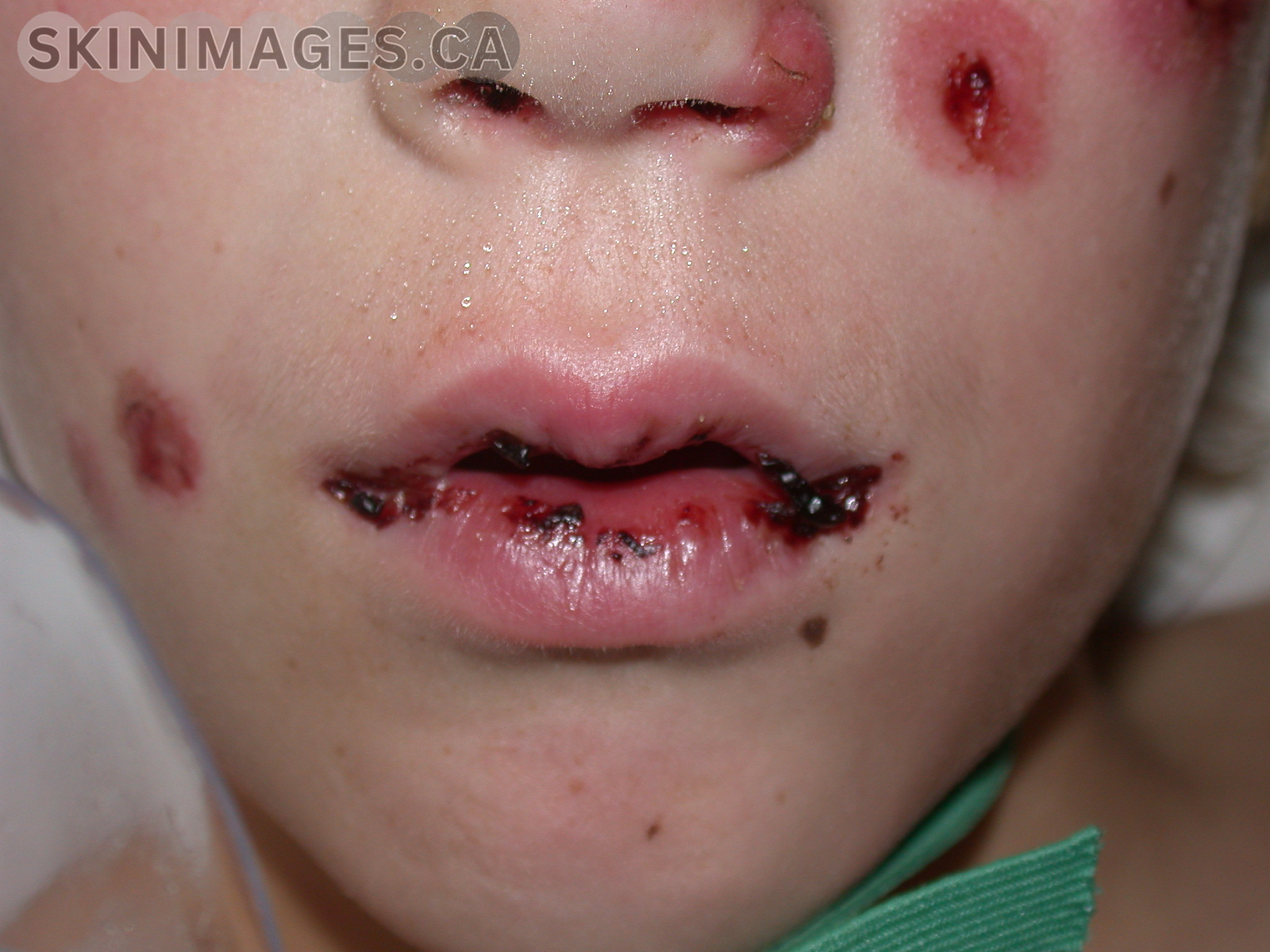
    Definition: The Herpes Simplex Virus (HSV) is a virus that causes herpes. Herpes is a common infection that can cause painful ulcers or blisters that may recur over time. The virus will remain dormant throughout a person’s life with the number of outbreaks decreasing with age. Two subtypes can cause infection: Herpes Simplex virus type 1 (HSV-1), and Herpes Simplex virus type 2 (HSV-2).

    Etiology: The HSV is a member of the Herpesvirales order of double-stranded DNA viruses. Both HSV subtypes affect the skin and mucous membranes. HSV is transmitted from oral or skin surfaces affected by the virus. Rarely, herpes can be transmitted vertically, causing neonatal herpes.

    While HSV-1 is associated with oral and facial infections, HSV-2 is associated with genital and rectal infections. Unique features of HSV include latency and reactivation. Following primary exposure there is a viral invasion of epithelial cells and intracellular replication at the site. HSV then ascends through the periaxonal sheath of sensory nerves to the host sacral ganglia- which acts as a reservoir for future outbreaks. As the virus replicates, it is dormant and can be reactivated by various stimuli.

    Epidemiology: As of 2016, the WHO estimates that 3.7 billion people under 50, (67% of the global population) had an HSV-1 infection, causing oral or genital herpes. It is estimated that 491 million people aged 15-49 worldwide were affected by HSV-2-causing genital herpes. Prevalence of HSV infection increases with age. The highest number of new infections is seen in adolescents. Women are affected by HSV-2 twice as often as men due to a more efficient route of sexual transmission.

    Signs: HSV infections are characterized by recurrent blisters that can rupture and become painful. Initial presentation can appear as painful genital ulcers, inguinal lymphadenopathy and fever. Areas that can become affected by clustered vesicles are the genitalia, perineum, buttocks, upper thigh, and perianal areas. The vesicles are small, umbilicated, and closely grouped with an erythematous base in recurrent herpes. They may have a zosteriform distribution when affecting the lower chest or lumbar region. In darker skin tones, scarring at the site of recurrent HSV attacks is more prominent and hyperpigmented. 

    Symptoms: Symptoms may vary between the initial outbreak and subsequent infections. Prodromal symptoms can appear up to 48 hours before an outbreak. These include fever, tender lymph nodes, myalgia, itching and tingling of the skin. Primary HSV-1 often presents as gingivostomatitis in children under 5 years. Primary HSV-2 usually presents as genital herpes following sexual activity.

    Differentials: Herpes Zoster, Syphilis, Cytomegalovirus, Chancroid, Hand foot and mouth disease

    Diagnosis: HSV diagnosis can be confirmed by culture or PCR of a viral swab taken from fresh vesicles.

    Treatment: HSV cannot be cured, so treatment is aimed at decreasing symptoms associated with first or recurrent episodes of herpes. Antiviral medication is commonly given in low daily doses to prevent outbreaks. NSAIDS can be given to help with pain related to sores.

    References:

    1. Cole S. Herpes Simplex Virus: Epidemiology, Diagnosis, and Treatment. Nurs Clin North Am. 2020;55(3):337-345. doi:10.1016/j.cnur.2020.05.004
    2. World Health Organization. Herpes simplex virus. Who.int. Published April 5, 2023. https://www.who.int/news-room/fact-sheets/detail/herpes-simplex-virus
    3. ‌Herpes simplex | DermNet NZ. dermnetnz.org. https://dermnetnz.org/topics/herpes-simplex
  • Pityriasis Rosea

    Pityriasis Rosea

    Pityriasis Rosea

    Pityriasis Rosea is an acute, self-limiting skin condition characterized by an initial herald patch followed by a widespread rash on the trunk and proximal extremities. It commonly affects individuals aged 10 to 35 years, with a slight female predominance. The exact cause is unknown, but it is believed to be associated with the reactivation of human herpesvirus 6 or 7.

    Clinically, the condition begins with a single large, scaly, pink patch (herald patch), typically on the chest, back, or abdomen. This is followed by smaller, oval, scaly patches that appear in a “Christmas tree” pattern. Some patients may experience mild prodromal symptoms such as headache, fever, or malaise before the rash appears. Diagnosis is primarily clinical, and possible differentials include tinea corporis, secondary syphilis, and psoriasis, among others. Treatment is usually unnecessary as the condition resolves on its own within 6–8 weeks. However, symptomatic management may include topical corticosteroids, oral antihistamines, and moisturizers to alleviate itching. The prognosis is excellent, with most cases resolving without complications. Recurrences are rare, but the condition can occasionally lead to prolonged skin discoloration​.

    References

    1. Villalon-Gomez JM. Pityriasis Rosea: Diagnosis and Treatment. Am Fam Physician. 2018;97(1):38-44.
    2. El-Hussein M, El-Tawil C, Nakhle R, Souaiby N. HHV 6-7 reactivation causing Pityriasis Rosea and labyrinthitis: A case report. Am J Emerg Med. 2020;38(9):1969.e1-1969.e3. doi:10.1016/j.ajem.2020.05.005
    3. Contreras-Ruiz J, Peternel S, Jiménez Gutiérrez C, Culav-Koscak I, Reveiz L, Silbermann-Reynoso ML. Interventions for pityriasis rosea. Cochrane Database Syst Rev. 2019;2019(10):CD005068. doi:10.1002/14651858.CD005068.pub3
  • Erythema Nodosum

    Erythema Nodosum

    Erythema Nodosum

    Erythema nodosum (EN) is an inflammatory condition characterized by the sudden appearance of painful, red nodules, typically on the shins. It is considered a hypersensitivity reaction to a variety of stimuli, including infections, medications, and systemic diseases.

    EN most commonly affects young adults, particularly women aged 20-40. The condition can be associated with infections such as streptococcal pharyngitis, tuberculosis, and fungal infections. It is also linked to systemic conditions like sarcoidosis, inflammatory bowel disease, and pregnancy. Certain medications, including oral contraceptives, can also trigger EN.

    Symptoms include the development of tender, erythematous nodules that usually measure between 1-5 cm in diameter. These nodules typically appear on the extensor surfaces of the lower legs but can also affect the thighs and forearms. The nodules evolve over several weeks, starting as firm and red, then becoming softer and less red before eventually resolving without ulceration or scarring.

    Treatment for erythema nodosum focuses on addressing the underlying cause and providing symptomatic relief. NSAIDs and potassium iodide can be used to reduce inflammation and pain. Bed rest and elevation of the affected limbs are recommended for severe cases. Identifying and treating any underlying infections or discontinuing causative medications are also crucial in managing EN.

    References:

    • Schwartz, R. A., Nervi, S. J., & Nervi, S. J. (2007). Erythema nodosum: A sign of systemic disease. American Family Physician, 75(5), 695-700.
    • Bohn, S., & Buchner, R. (2006). Erythema nodosum: Etiology, diagnosis, and treatment. Journal of the American Academy of Dermatology, 55(6), 1133-1146.
    • Mert, A., Kumbasar, H., Ozaras, R., Erten, S., Tasdelen, F., & Bilir, M. (2007). Erythema nodosum: An evaluation of 100 cases. Clinical and Experimental Rheumatology, 25(4), 563-570.
  • Erythema Multiforme

    Erythema Multiforme

    Erythema Multiforme

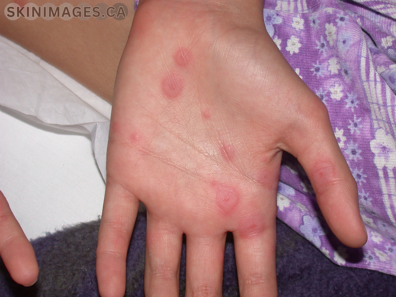
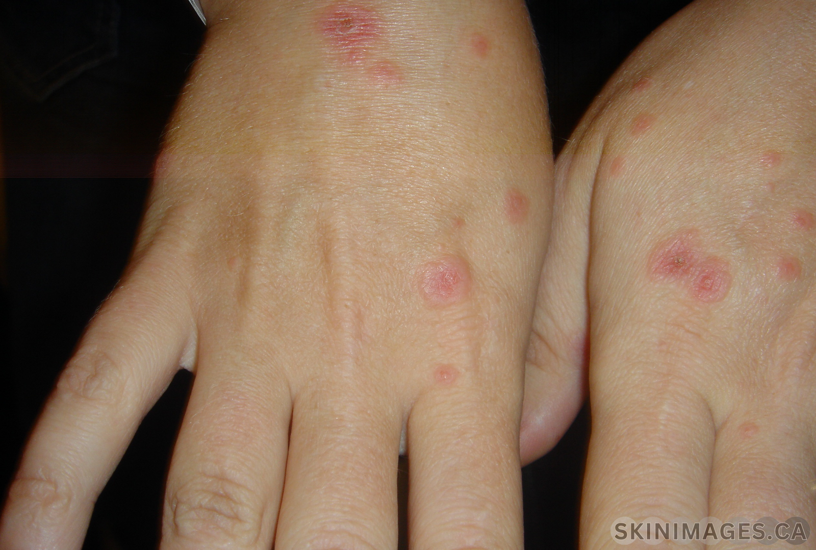
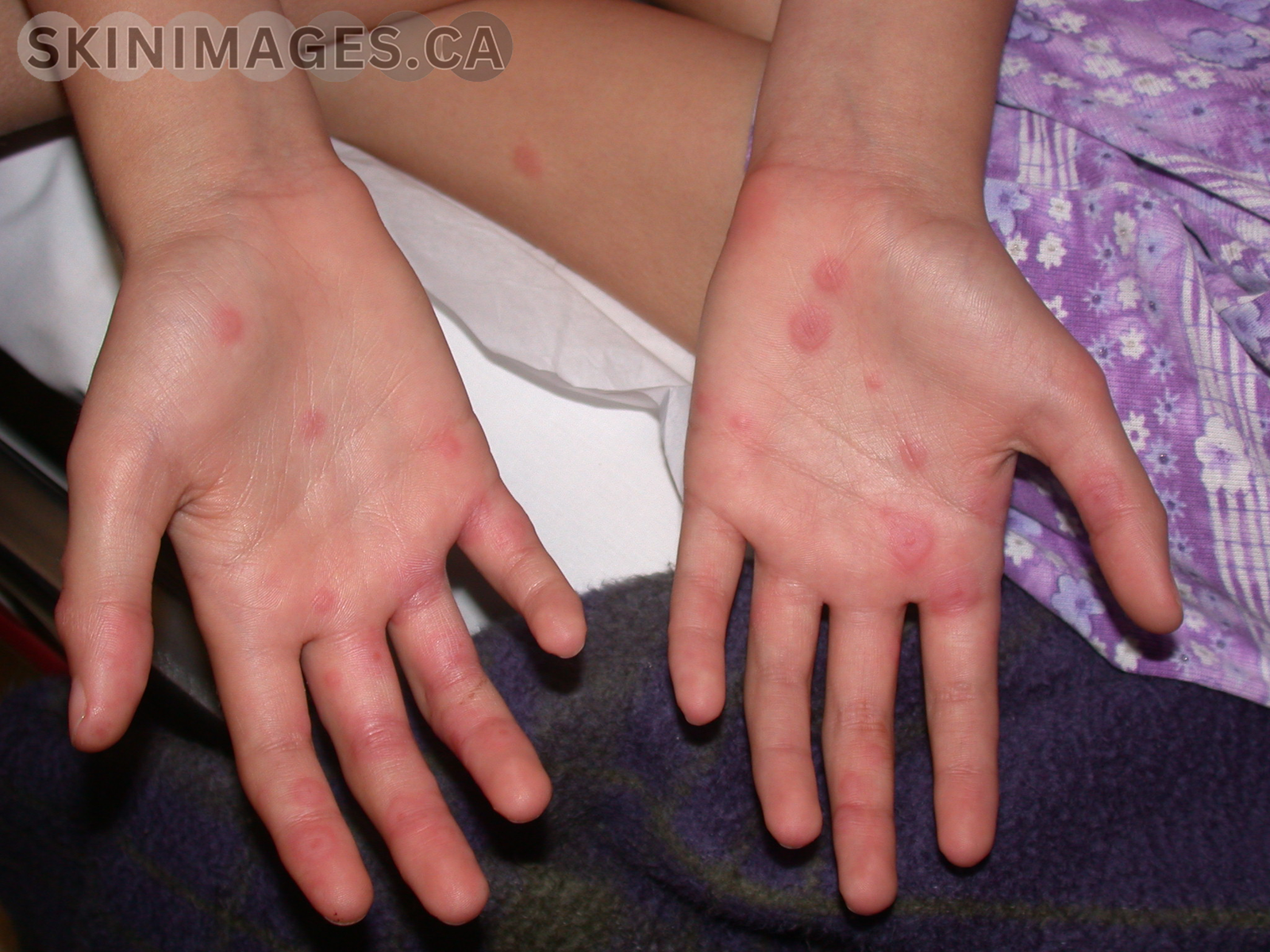
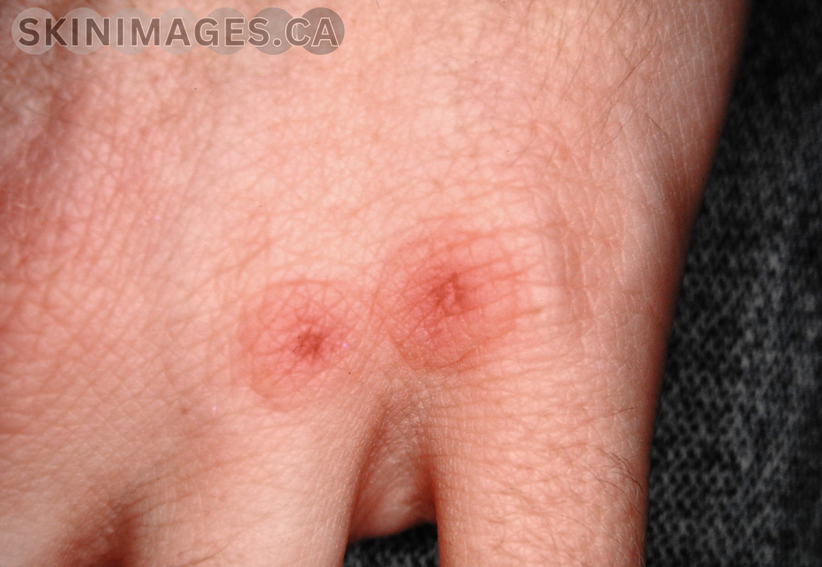
    Erythema multiforme (EM) is an acute, self-limiting, hypersensitivity reaction characterized by target-like lesions on the skin and mucous membranes. It is most commonly triggered by infections, particularly herpes simplex virus (HSV), but can also be associated with medications and other factors.

    EM predominantly affects young adults, with a slight male predominance. The condition is classified into two types: erythema multiforme minor and erythema multiforme major. EM minor typically presents with symmetrical, red, target-like lesions primarily on the extremities, while EM major includes mucosal involvement and more extensive skin lesions.

    EM symptoms include the sudden onset of erythematous macules, papules, and target lesions that may be pruritic or painful. In EM major, mucosal involvement can lead to painful erosions and ulcerations in the oral cavity, genitalia, and ocular regions.

    Treatment for EM focuses on identifying and removing the underlying cause. Supportive care includes the use of topical corticosteroids, antihistamines, and analgesics to manage symptoms. Severe cases of EM major may require systemic corticosteroids and hospitalization for supportive care. Preventive antiviral therapy may be considered in recurrent cases associated with HSV.

    References:

    • Trayes, K. P., Love, G., & Studdiford, J. S. (2019). Erythema multiforme: Recognition and management. American Family Physician, 100(2), 82-88.
    • Lerch, M., & Mainetti, C. (2018). Erythema multiforme. Journal of the European Academy of Dermatology and Venereology, 32(5), 781-788.
    • Wetter, D. A., & Davis, M. D. (2010). Recurrent erythema multiforme: Clinical characteristics, etiologic associations, and treatment in a series of 48 patients at Mayo Clinic, 2000 to 2007. Journal of the American Academy of Dermatology, 62(1), 45-53.