Body Part: Head

  • Alopecia Areata

    Alopecia Areata

    Alopecia Areata

    Definition: A common autoimmune condition that is marked by abrupt, non-scarring hair loss on the scalp and other parts of the body go (1)

    Etiology: Many etiologies have been proposed but it is believed that the condition is caused by an autoimmune attack on the hair follicles, leading to hair loss. In addition, environmental factors, genetic predisposition and stress may also play a role (1,2).

    Epidemiology: Alopecia Areat (AA) affects 0.1-0.2% of the overall population with no gender predilection. (1,2).

    Signs: AA is characterized by well-demarcated oval or round patches of hair loss, the lesions may be solitary or multiple with 90% of cases affecting the scalp (2).

    Symptoms: Typically asymptomatic however some patients may experience nail changes, tingling or burning sensation, irritated eyes if loss of hair in eyelashes or eyebrows (3).

    Differentials: Telogen effluvium, Trichotillomania,Tinea capitis, Early scarring Alopecia, Anagen effluvium, Systemic lupus erythematosus, Secondary syphilis and androgenic Alopecia (1,2). 

    Diagnosis: The diagnosis is clinical with a thorough history and routine investigations such as complete hemogram, anemia panel, erythrocyte sedimentation rate, etc (2). Dermoscopic histological examination is a great tool in diagnosing AA and in more uncertain cases a biopsy may also be performed (1).

    Treatment: In a majority of cases the condition is self-limited and regrowth typically will happen within a year (1). In patients where AA is persistent corticosteroids can be prescribed to stimulate hair growth follow by minoxidil to maintain the growth (3).

    References: (AMA)

    1.     Khan Mohammad Beigi P. Alopecia areata. Published online 2018. doi:10.1007/978-3-319-72134-7 

    1. Amin SS, Sachdeva S. Alopecia areata: A Review. Journal of the Saudi Society of Dermatology & Dermatologic Surgery. 2013;17(2):37-45. doi:10.1016/j.jssdds.2013.05.004 
    2. Ludmann P. Hair loss types: Alopecia areata diagnosis and treatment. American Academy of Dermatology. 2023. Accessed August 18, 2024. https://www.aad.org/public/diseases/hair-loss/types/alopecia/treatment. 
  • Herpes Zoster

    Herpes Zoster

    Herpes Zoster

    Definition: Herpes zoster, also known as shingles, is a localized, painful, blistering rash that is caused by a reactivation of the varicella-zoster virus.

    Etiology: The varicella-zoster virus (VZV) is a member of the Herpesvirales order of double-stranded DNA viruses, that causes chickenpox. The VZV establishes latency in the dorsal root ganglia nerve cells of the spinal cord after primary infection. Once reactivated, the virus replicates in neuronal cell bodies and migrates down sensory nerves to the skin, causing herpes zoster. Pain associated with zoster is due to inflamed nerves that are affected by the virus. The virus can be transmitted via skin-to-skin contact or droplet inhalation.

    Epidemiology: The incidence of herpes zoster is 1.2- 3.4 per 1000 persons per year in individuals younger than 65 years. In individuals older than 65 years, the incidence is 3.9- 11.8  per 1000 persons per year. Recurrences are often seen in immunocompromised patients. 

    Signs: The dermatological involvement of herpes zoster is centripetal and follows a dermatomal distribution pattern. This is typically unilateral and may vary depending on the affected dermatome, age and health of the patient. Following the onset of pain, the rash that erupts is a crop of red papules that is painful, erythematous and blistering. On dark skin, the “shingles band” may appear as gray, dark brown or violaceous papules. The rash then develops forming vesicles which can cause discolouration of the surrounding skin depending on skin tone.The lesions continue to erupt and become pustular and crust over. 

    Symptoms: Early symptoms include localized or widespread pain without tenderness or skin changes. The zoster rash is painful within the associated area of skin. Individuals may feel unwell with a fever and headache. In uncomplicated cases, pain and general symptoms subside as the eruption disappears.

    Differentials: Herpes simplex, dermatitis herpetiformis, impetigo, contact dermatitis, candidiasis.

    Diagnosis: Herpes zoster is diagnosed clinically with pain, characteristic morphology and dermatomal distribution. The rash may form a pattern called the zosteriform herpes simplex. Diagnostic tests include direct fluorescent antibody, PCR and Tzanck smear of vesicular fluid or blood.  

    Treatment: Several antiviral drugs are available to treat shingles such as acyclovir and valacyclovir. They shorten the length and severity of illness. Over-the-counter pain relief medication can also aid in the pain caused by shingles. The CDC recommends 2 doses of the recombinant zoster vaccine to prevent shingles and its complications in individuals 50 years and older. Vaccination is also recommended in younger, immunocompromised patients.

    References:

    1. Nair PA, Patel BC. Herpes Zoster. In: StatPearls. Treasure Island (FL): StatPearls Publishing; September 4, 2023.
    2. Ayoade F, Kumar S. Varicella-Zoster Virus (Chickenpox). In: StatPearls. Treasure Island (FL): StatPearls Publishing; October 15, 2022.
    3. Herpes zoster | DermNet NZ. dermnetnz.org. https://dermnetnz.org/topics/herpes-zoster
    4. ‌CDC. Shingles. Centers for Disease Control and Prevention. Published 2019. https://www.cdc.gov/shingles/about/index.html
  • Herpes Zoster

    Herpes Zoster

    Herpes Zoster

    Definition: Herpes zoster, also known as shingles, is a localized, painful, blistering rash that is caused by a reactivation of the varicella-zoster virus.

    Etiology: The varicella-zoster virus (VZV) is a member of the Herpesvirales order of double-stranded DNA viruses, that causes chickenpox. The VZV establishes latency in the dorsal root ganglia nerve cells of the spinal cord after primary infection. Once reactivated, the virus replicates in neuronal cell bodies and migrates down sensory nerves to the skin, causing herpes zoster. Pain associated with zoster is due to inflamed nerves that are affected by the virus. The virus can be transmitted via skin-to-skin contact or droplet inhalation.

    Epidemiology: The incidence of herpes zoster is 1.2- 3.4 per 1000 persons per year in individuals younger than 65 years. In individuals older than 65 years, the incidence is 3.9- 11.8  per 1000 persons per year. Recurrences are often seen in immunocompromised patients. 

    Signs: The dermatological involvement of herpes zoster is centripetal and follows a dermatomal distribution pattern. This is typically unilateral and may vary depending on the affected dermatome, age and health of the patient. Following the onset of pain, the rash that erupts is a crop of red papules that is painful, erythematous and blistering. On dark skin, the “shingles band” may appear as gray, dark brown or violaceous papules. The rash then develops forming vesicles which can cause discolouration of the surrounding skin depending on skin tone.The lesions continue to erupt and become pustular and crust over. 

    Symptoms: Early symptoms include localized or widespread pain without tenderness or skin changes. The zoster rash is painful within the associated area of skin. Individuals may feel unwell with a fever and headache. In uncomplicated cases, pain and general symptoms subside as the eruption disappears.

    Differentials: Herpes simplex, dermatitis herpetiformis, impetigo, contact dermatitis, candidiasis.

    Diagnosis: Herpes zoster is diagnosed clinically with pain, characteristic morphology and dermatomal distribution. The rash may form a pattern called the zosteriform herpes simplex. Diagnostic tests include direct fluorescent antibody, PCR and Tzanck smear of vesicular fluid or blood.  

    Treatment: Several antiviral drugs are available to treat shingles such as acyclovir and valacyclovir. They shorten the length and severity of illness. Over-the-counter pain relief medication can also aid in the pain caused by shingles. The CDC recommends 2 doses of the recombinant zoster vaccine to prevent shingles and its complications in individuals 50 years and older. Vaccination is also recommended in younger, immunocompromised patients.

    References:

    ‌CDC. Shingles. Centers for Disease Control and Prevention. Published 2019. https://www.cdc.gov/shingles/about/index.html

    Nair PA, Patel BC. Herpes Zoster. In: StatPearls. Treasure Island (FL): StatPearls Publishing; September 4, 2023.

    Ayoade F, Kumar S. Varicella-Zoster Virus (Chickenpox). In: StatPearls. Treasure Island (FL): StatPearls Publishing; October 15, 2022.

    Herpes zoster | DermNet NZ. dermnetnz.org. https://dermnetnz.org/topics/herpes-zoster

  • Acne, Hormonal with Hirsutism

    Acne, Hormonal with Hirsutism

    Acne, Hormonal with Hirsutism

    Definition: Hormonal acne with Hirsutism is a subtype of acne often linked with androgen excess. Androgens increase sebum production and follicular keratosis, which are key factors in the development of acne. Hirsutism is defined as the excessive growth of terminal hair in women in a pattern typically seen in men.

    Epidemiology: This condition frequently affects women from their late teens into adulthood and is often linked to polycystic ovary syndrome (PCOS).

    Signs: Along with typical acne lesions (comedones, papules, pustules), patients may show signs of hirsutism, such as excessive facial and body hair, and other indications of hyperandrogenism like alopecia and irregular menstruation.

    Symptoms: Patients may experience symptoms of acne, such as inflammation and tenderness, along with hirsutism. These issues can lead to psychological distress, driven by cosmetic concerns and social stigma.

    Differentials: Other types of hyperandrogenic disorders such as congenital adrenal hyperplasia, androgen-secreting tumours, and other types of acne.

    Diagnosis: A comprehensive history and targeted physical examination are crucial for evaluating androgen excess in the clinic. This includes a clinical assessment of acne and hirsutism, supported by laboratory tests to measure androgen levels, and imaging studies to evaluate ovarian morphology if PCOS is suspected.

    Treatment: Treatment for acne includes topical anti-acne agents, oral antibiotics like tetracycline, antiandrogens (such as hormone therapy and birth control pills), and oral isotretinoin. Management of hirsutism typically involves a dual approach: pharmacological therapy to reduce androgen secretion and/or action, and the removal of existing terminal hair.

    References: (AMA)

    1. Lumezi BG, Pupovci HL, Berisha VL, Goçi AU, Gerqari A. Acne in hirsute women. Postepy Dermatol Alergol. 2014;31(6):356-361. doi:10.5114/pdia.2014.47118
    2. Yildiz BO. Diagnosis of hyperandrogenism: clinical criteria. Best Practice & Research Clinical Endocrinology & Metabolism. 2006;20(2):167-176. doi:https://doi.org/10.1016/j.beem.2006.02.004
    3. Tewary S, Davies R, Prakash A. Hirsutism. Obstetrics, Gynaecology & Reproductive Medicine. Published online March 12, 2021. doi:https://doi.org/10.1016/j.ogrm.2021.02.004
  • Acne Keloidalis Nuchae

    Acne Keloidalis Nuchae

    Acne Keloidalis Nuchae

    Definition: Acne keloidalis nuchae (AKN) also known as folliculitis keloidalis nuchae (FKN) is a chronic debilitating condition that is characterized by keloid-life plaques that usually affects the occipital area or nape of the neck. Untreated AKN can lead to scarring folliculitis.

    Epidemiology: Acne keloidalis nuchae is thought to predominantly affect men of African American descent but a few studies have shown it to significantly affect other ethnicities. It is seen in post-pubertal males and is uncommon beyond the age of 55.

    Signs: Firm, flesh-coloured, pink or flesh-coloured papules and nodules may aggregate into sizable plaques resembling keloid. In the afflicted locations, these lesions frequently result in permanent hair loss and scarring.

    Symptoms: Symptoms typically develop from a haircut or irritation from wearing a sports helmet and tend to appear a few hours to days after the irritation. In active lesions, pruritus, discomfort, and contact bleeding are frequent which may cause men to avoid haircuts.

    Differentials: AKN is often mistaken for a keloidal condition and shares some features with cicatricial alopecia.

    Diagnosis: Based mostly on the location and distinctive look of the lesions, the diagnosis is made clinically. In rare circumstances, a biopsy may be done to rule out other disorders.

    Treatment: Treatment options include topical and intralesional corticosteroids, topical retinoids, topical antibacterials, oral antibiotics for secondary infections, and laser therapy. In refractory cases, surgical excision or radiation therapy may be considered as well as oral isotretinoin.

    References: (AMA)

    1. Umar S, Lee DJ, Lullo JJ. A Retrospective Cohort Study and Clinical Classification System of Acne Keloidalis Nuchae. J Clin Aesthet Dermatol. 2021;14(4):E61-E67.
    2. Ogunbiyi A. Acne keloidalis nuchae: prevalence, impact, and management challenges. Clin Cosmet Investig Dermatol. 2016;9:483-489. Published 2016 Dec 14. doi:10.2147/CCID.S99225
    3. Maranda EL, Simmons BJ, Nguyen AH, Lim VM, Keri JE. Treatment of Acne Keloidalis Nuchae: A Systematic Review of the Literature. Dermatol Ther (Heidelb). 2016;6(3):363-378. doi:10.1007/s13555-016-0134-5
  • Erythema Multiforme

    Erythema Multiforme

    Erythema Multiforme

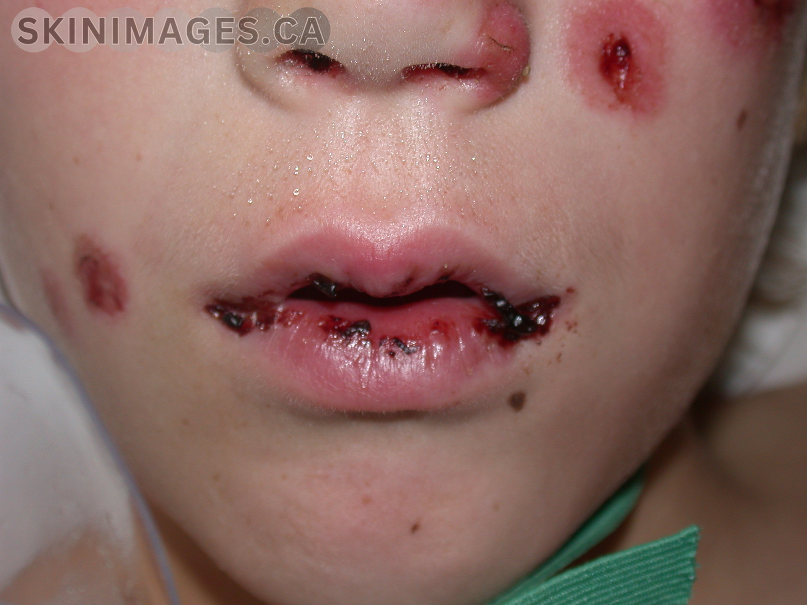
    Erythema multiforme (EM) is an acute, self-limiting, hypersensitivity reaction characterized by target-like lesions on the skin and mucous membranes. It is most commonly triggered by infections, particularly herpes simplex virus (HSV), but can also be associated with medications and other factors.

    EM predominantly affects young adults, with a slight male predominance. The condition is classified into two types: erythema multiforme minor and erythema multiforme major. EM minor typically presents with symmetrical, red, target-like lesions primarily on the extremities, while EM major includes mucosal involvement and more extensive skin lesions.

    EM symptoms include the sudden onset of erythematous macules, papules, and target lesions that may be pruritic or painful. In EM major, mucosal involvement can lead to painful erosions and ulcerations in the oral cavity, genitalia, and ocular regions.

    Treatment for EM focuses on identifying and removing the underlying cause. Supportive care includes the use of topical corticosteroids, antihistamines, and analgesics to manage symptoms. Severe cases of EM major may require systemic corticosteroids and hospitalization for supportive care. Preventive antiviral therapy may be considered in recurrent cases associated with HSV.

    References:

    • Trayes, K. P., Love, G., & Studdiford, J. S. (2019). Erythema multiforme: Recognition and management. American Family Physician, 100(2), 82-88.
    • Lerch, M., & Mainetti, C. (2018). Erythema multiforme. Journal of the European Academy of Dermatology and Venereology, 32(5), 781-788.
    • Wetter, D. A., & Davis, M. D. (2010). Recurrent erythema multiforme: Clinical characteristics, etiologic associations, and treatment in a series of 48 patients at Mayo Clinic, 2000 to 2007. Journal of the American Academy of Dermatology, 62(1), 45-53.
  • Hemangioma

    Hemangioma

    Hemangioma

    Definition: A hemangioma is a benign proliferative blood vessel neoplasm. Hemangiomas can arise early or later in life, and there are several hemangiomas. The most common type is a cherry angioma. Congenital hemangiomas are present at birth while infantile hemangiomas appear later within the first few weeks of life.

    Etiology: Hemangiomas are caused by endothelial cell proliferation. Studies suggest this may occur in three ways: increased GLUT- 1 and VEGF expression, stem cells originating from trophoblasts or de novo formation.

    Epidemiology: Hemangiomas are the most common benign tumours of infancy, affecting approximately 4-5% of newborns. Infantile hemangiomas appear more frequently in Caucasian infants. Premature infants, infants with elder mothers and low birth weight infants are also more susceptible to developing hemangiomas. Most cases of hemangiomas are sporadic but are associated with an autosomal dominant inheritance pattern with no specific gene implicated.

    Signs: Cherry hemangiomas typically appear as multiple firm erythematous or violaceous papules less than 1 cm in diameter. They can be scattered across any body surface If thrombosed, they can appear darker in colour before close dermatoscopic inspection when the red or purple colour is more easily seen. Infantile hemangiomas are usually solitary and appear on the head and neck. Clinical features are determined by the lesion’s depth, distribution pattern (usually facial in 77% of cases) and growth phases.

    Symptoms: Cherry angiomas may be asymptomatic. Superficial, deep and mixed infantile hemangiomas are usually non-tender.

    Differentials: Port-wine stain, Congenital hemangioma, Angiokeratoma, Pyogenic granuloma, Kaposiform hemangioendothelioma

    Diagnosis: Hemangiomas are diagnosed clinically. A skin biopsy may also be performed. Infantile hemangiomas are positive for GLUT-1 staining. Ultrasound, CT and MRI may be performed to determine the depth of infantile hemangiomas where there are no superficial skin changes.

    Treatment: Cherry angiomas are typically harmless and are left untreated. In some cases, they must be excised to rule out nodular melanoma.  Infantile hemangiomas may resolve on their own. Complicated infantile hemangiomas may require treatment. This is in the form of beta-blockers, oral prednisone, and topical corticosteroids.

    References:

    1. Infantile haemangioma: Definition and pathogenesis | DermNet NZ. dermnetnz.org. https://dermnetnz.org/topics/infantile-haemangioma-definition-and-pathogenesis
    2. Chamli A, Aggarwal P, Jamil RT, Litaiem N. Hemangioma. In: StatPearls. Treasure Island (FL): StatPearls Publishing; June 12, 2023.
    3. ‌Angiomas. Spider naevus | DermNet NZ. dermnetnz.org. https://dermnetnz.org/topics/cherry-angioma
    4. Dicko A, Safi T, Tounkara TM, et al. Prevalence des hemangiomes infantiles sur peau noire au Mali [Prevalence of infantile haemangiomas on black skin]. Mali Med. 2017;32(4):18-20.