Body Part: Hand

  • Porphyria Cutanea Tarda

    Porphyria Cutanea Tarda

    Porphyria Cutanea Tarda

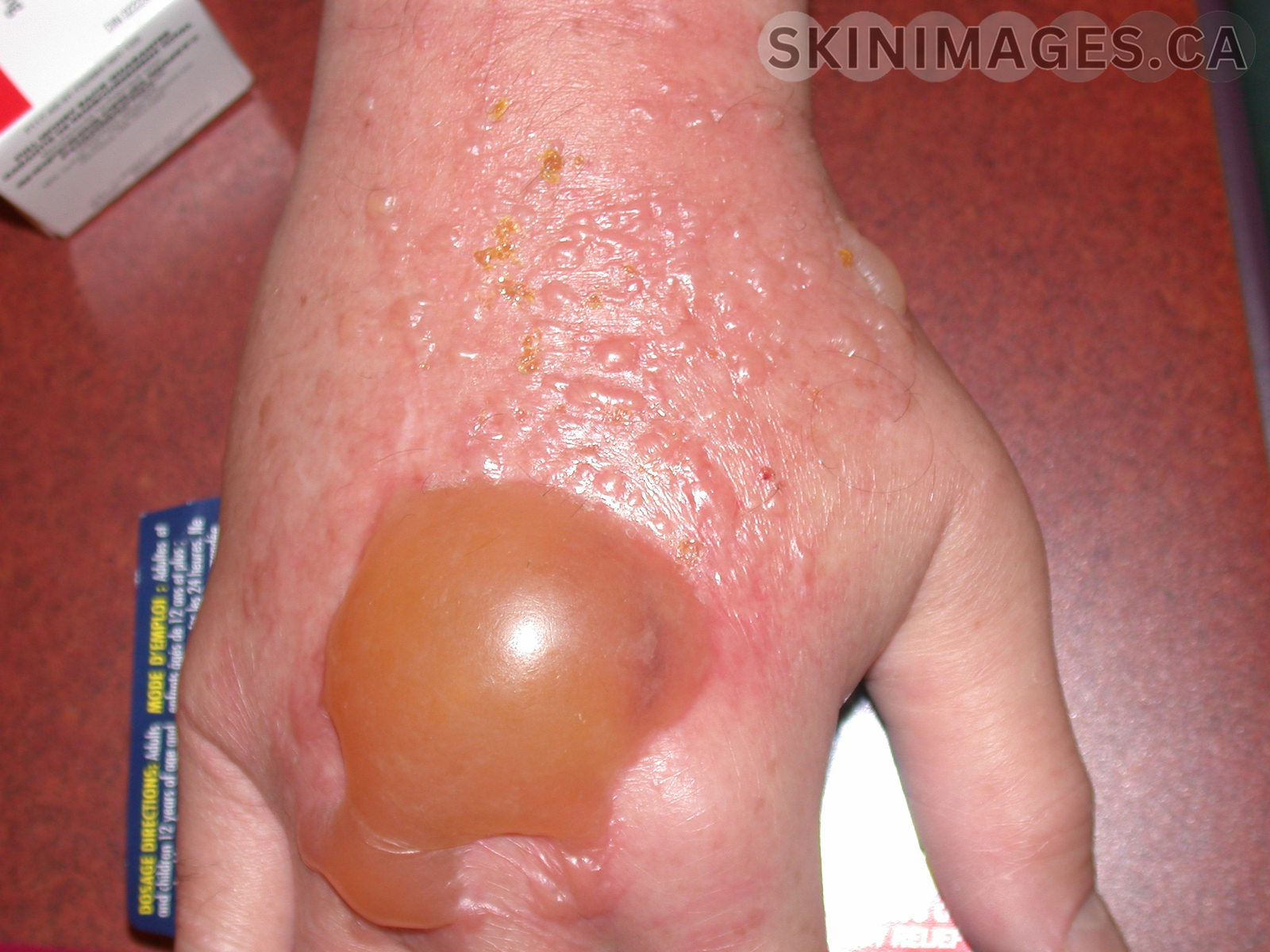
    Condition Name: Porphyria Cutanea Tarda (PCT)

    Definition: PCT is a disorder characterized by blistering lesions on sun-exposed skin due to a deficiency of the enzyme uroporphyrinogen decarboxylase (UROD).

    Etiology: PCT can be inherited or acquired. Acquired PCT is often linked to liver disease, such as hepatitis C, alcohol abuse, or hemochromatosis, while familial PCT involves a genetic mutation in the UROD gene.

    Epidemiology: PCT is the most common type of porphyria, typically presenting in adults in their 40s or 50s. Familial cases tend to present earlier in life, while sporadic cases often appear in the fourth or fifth decades.

    Signs: Blistering, fragile skin on sun-exposed areas like the hands and face, with potential hyperpigmentation, scarring, and milia.

    Symptoms: Skin sensitivity to sunlight, pain and discomfort in affected areas, and dark urine due to elevated porphyrins.

    Differentials: Variegate porphyria, bullous pemphigoid, lupus erythematosus.

    Diagnosis: Confirmed through porphyrin level testing in blood, urine, and stool, with a Wood’s lamp examination revealing fluorescence in urine.

    Treatment: Therapeutic phlebotomy to reduce iron levels, low-dose antimalarials like hydroxychloroquine, and strict sun protection.

    References:

    1.     Singal AK. Porphyria cutanea tarda: Recent update. Mol Genet Metab. 2019;128(3):271-281. doi:10.1016/j.ymgme.2019.01.004

    2.     Frank J, Poblete-Gutiérrez P. Porphyria cutanea tarda–when skin meets liver. Best Pract Res Clin Gastroenterol. 2010;24(5):735-745. doi:10.1016/j.bpg.2010.07.002

  • Granuloma Annulare

    Granuloma Annulare

    Granuloma annulare 

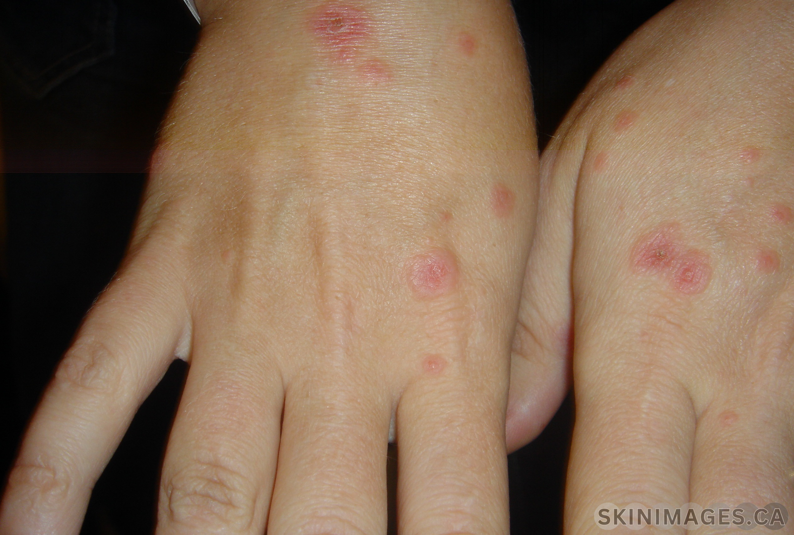
    Condition Name: Granuloma annulare

    Definition: Granuloma annulare is a benign, chronic skin condition characterized by the formation of annular plaques or papules.

    Etiology: The exact cause of granuloma annulare is unknown, though it is believed to involve an immune-mediated response.

    Epidemiology: Granuloma annulare affects individuals of all ages but is more commonly seen in children and young adults. It is slightly more prevalent in females than in males. The prevalence in the general population is relatively low, and the condition is not typically associated with significant morbidity.

    Signs: The appearance of small, firm, flesh-colored or erythematous papules that coalesce to form rings with a central depression. These lesions most commonly appear on the hands, feet, elbows, and knees but can occur on any part of the body.

    Symptoms: The lesions are usually asymptomatic but can occasionally cause mild itching.

    Differentials: Conditions such as tinea corporis, sarcoidosis, and necrobiosis lipoidica should be considered.

    Diagnosis: Diagnosis is primarily clinical, based on the characteristic appearance of the lesions. A skin biopsy can confirm the diagnosis by revealing necrobiotic collagen surrounded by histiocytes and multinucleated giant cells.

    Treatment: Treatment is often unnecessary as granuloma annulare can resolve spontaneously, particularly in localized cases. Treatment options for persistent or widespread cases include topical or intralesional corticosteroids, cryotherapy, or laser therapy. Systemic treatments, such as dapsone, isotretinoin, or hydroxychloroquine, may be considered for more severe cases.

    References:

    ●       Piette, E. W., & Rosenbach, M. (2016). Granuloma annulare: Pathogenesis, disease associations, and triggers, and therapeutic options. Journal of the American Academy of Dermatology, 75(3), 467-479. doi:10.1016/j.jaad.2016.02.1222

    ●       Marneros, A. G., & Bruckner, A. L. (2010). “Granuloma annulare.” Journal of the American Academy of Dermatology, 62(2), 207-222. doi:10.1016/j.jaad.2009.03.044

  • Allergic Contact Dermatitis

    Allergic Contact Dermatitis

    Allergic Contact Dermatitis 

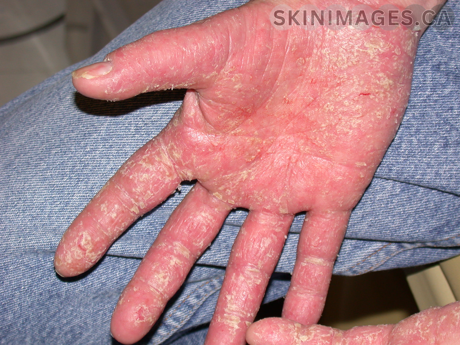
    Definition: Allergic contact dermatitis (ACD) is a skin condition that occurs when the skin comes into contact with an allergen, leading to an immune response and subsequent inflammation(1).

    Etiology: ACD is caused by an allergic reaction to substances that come into direct contact with the skin. Common allergens include nickel, fragrances, preservatives, latex, and certain plants like poison ivy (1,3). The immune system recognizes these substances as harmful, triggering an inflammatory response(1).

    Epidemiology: ACD affects individuals of all ages, but it is more prevalent in adults due to cumulative exposure to allergens over time. It is common with some studies demonstrating a prevalence rate of 20% in the general population (2). It is particularly common in certain occupations that involve frequent contact with irritants and allergens, such as healthcare workers, hairdressers, and construction workers (2). 

    Signs: Key signs of ACD include erythema, edema, vesicles, and lichenification in chronic cases (1). These signs typically appear in areas directly exposed to the allergen(1,2).

    Symptoms: Symptoms of ACD include intense itching, burning, and discomfort at the site of contact(3). The skin may also become dry, cracked, and scaly if exposure continues or if the dermatitis becomes chronic(3)

    Differentials: Differential diagnoses for ACD include irritant contact dermatitis, atopic dermatitis, psoriasis, fungal infections, and seborrheic dermatitis (1).

    Diagnosis: Diagnosis of ACD is primarily clinical, based on the history of exposure and characteristic skin findings(1). Patch testing is the gold standard and is used to identify the allergens that cause allergic contact dermatitis (1).

    Treatment: The cornerstone of ACD treatment is identifying and avoiding the offending allergen(1). Topical corticosteroids are commonly prescribed to reduce inflammation and alleviate symptoms(1). In severe cases, systemic corticosteroids may be required. Avoidance necessitates thorough label-checking for the allergen and related cross-reactive substances. Understanding these cross-reactive ingredients and knowing where a particular allergen is commonly found can aid in effectively advising the patient (1).

    References: (AMA)

    1. Kanwaljit K. Brar MD, AbstractObjectiveTo familiarize the reader with the mechanisms and causes of contact dermatitis.Data SourcesRecent research articles, Dhingra N, et al. A review of contact dermatitis. Annals of Allergy, Asthma & Immunology. October 20, 2020. Accessed August 10, 2024. https://www.sciencedirect.com/science/article/pii/S1081120620310802.
    2. Stacy Nassau MD, Jacob SE, Warshaw EM, et al. Allergic contact dermatitis. Medical Clinics of North America. October 28, 2019. Accessed August 10, 2024. https://www.sciencedirect.com/science/article/pii/S0025712519300884. 
    3. Brandon L. Adler M. Allergic contact dermatitis. JAMA Dermatology. March 1, 2021. Accessed August 10, 2024. https://jamanetwork.com/journals/jamadermatology/article-abstract/2775575. 
  • Erythema Multiforme

    Erythema Multiforme

    Erythema Multiforme

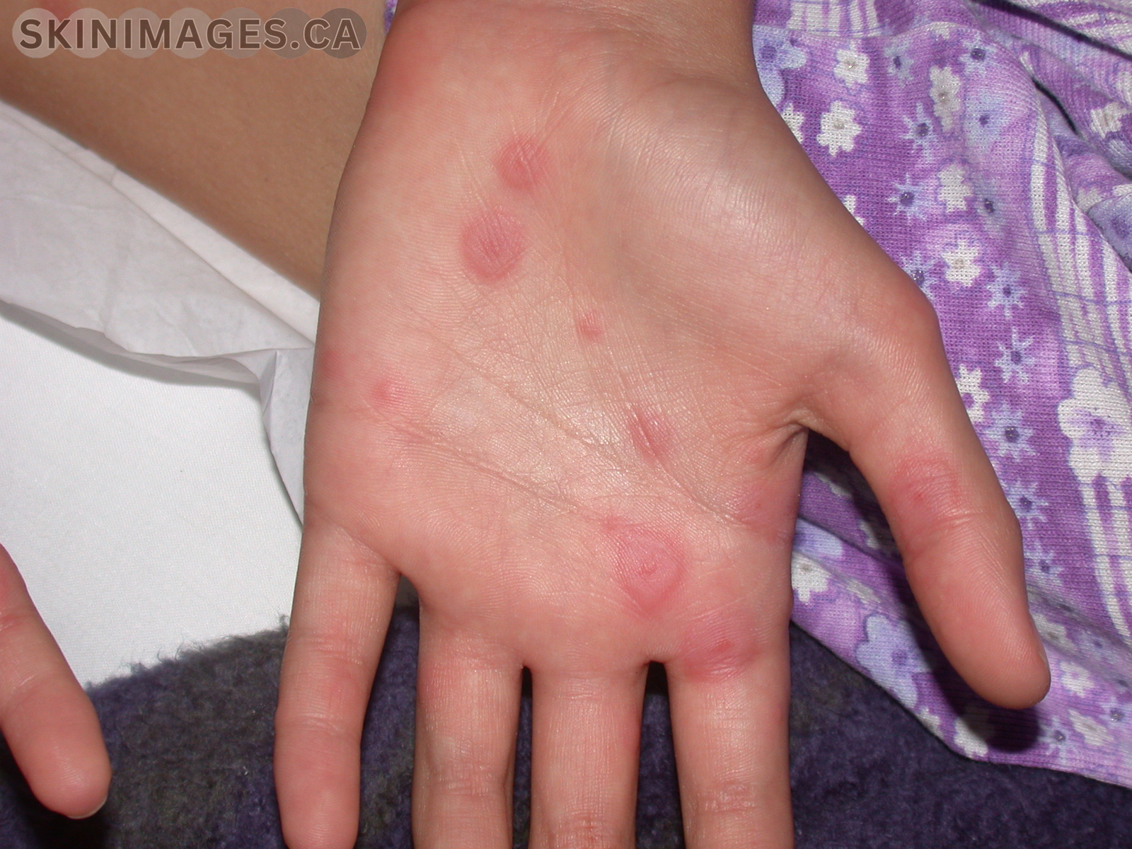
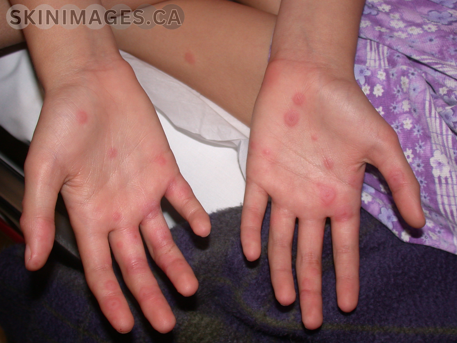
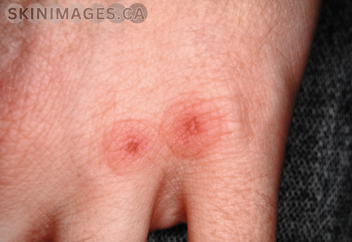
    Erythema multiforme (EM) is an acute, self-limiting, hypersensitivity reaction characterized by target-like lesions on the skin and mucous membranes. It is most commonly triggered by infections, particularly herpes simplex virus (HSV), but can also be associated with medications and other factors.

    EM predominantly affects young adults, with a slight male predominance. The condition is classified into two types: erythema multiforme minor and erythema multiforme major. EM minor typically presents with symmetrical, red, target-like lesions primarily on the extremities, while EM major includes mucosal involvement and more extensive skin lesions.

    EM symptoms include the sudden onset of erythematous macules, papules, and target lesions that may be pruritic or painful. In EM major, mucosal involvement can lead to painful erosions and ulcerations in the oral cavity, genitalia, and ocular regions.

    Treatment for EM focuses on identifying and removing the underlying cause. Supportive care includes the use of topical corticosteroids, antihistamines, and analgesics to manage symptoms. Severe cases of EM major may require systemic corticosteroids and hospitalization for supportive care. Preventive antiviral therapy may be considered in recurrent cases associated with HSV.

    References:

    • Trayes, K. P., Love, G., & Studdiford, J. S. (2019). Erythema multiforme: Recognition and management. American Family Physician, 100(2), 82-88.
    • Lerch, M., & Mainetti, C. (2018). Erythema multiforme. Journal of the European Academy of Dermatology and Venereology, 32(5), 781-788.
    • Wetter, D. A., & Davis, M. D. (2010). Recurrent erythema multiforme: Clinical characteristics, etiologic associations, and treatment in a series of 48 patients at Mayo Clinic, 2000 to 2007. Journal of the American Academy of Dermatology, 62(1), 45-53.